Also found in: Dictionary, Thesaurus, Medical, Encyclopedia.
| Diencephalon | |
|---|---|
 | |
| Details | |
| Precursor | Prosencephalon, derived from the neural tube |
| Part of | Human brain |
| Parts | Thalamus, the hypothalamus, the epithalamus and the subthalamus |
| Identifiers | |
| Latin | diencephalon |
| MeSH | D004027 |
| NeuroLex ID | birnlex_1503 |
| TA98 | A14.1.03.007 A14.1.08.001 |
| TA2 | 5661 |
| TH | H3.11.03.5.00001 |
| FMA | 62001 |
| Anatomical terms of neuroanatomy | |
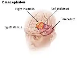
The diencephalon is a division of the forebrain (embryonic prosencephalon), and is situated between the telencephalon and the midbrain (embryonic mesencephalon). The diencephalon has also been known as the 'tweenbrain in older literature.[1] It consists of structures that are on either side of the third ventricle, including the thalamus, the hypothalamus, the epithalamus and the subthalamus.
The diencephalon is one of the main vesicles of the brain formed during embryogenesis. During the third week of development a neural tube is created from the ectoderm, one of the three primary germ layers. The tube forms three main vesicles during the third week of development: the prosencephalon, the mesencephalon and the rhombencephalon. The prosencephalon gradually divides into the telencephalon and the diencephalon.
Structure
The diencephalon consists of the following structures:
- Thalamus
- Hypothalamus including the posterior pituitary
- Epithalamus which consists of:
- Anterior and Posterior Paraventricular nuclei
- Medial and lateral habenular nuclei
- Stria medullaris thalami
- Posterior commissure
- Pineal body
- Subthalamus
Attachments
The optic nerve (CNII) attaches to the diencephalon. The optic nerve is a sensory (afferent) nerve responsible for vision and sight ; it runs from the eye through the optic canal in the skull and attaches to the diencephalon. The retina itself is derived from the optic cup, a part of the embryonic diencephalon.
Function
The diencephalon is the region of the embryonic vertebrate neural tube that gives rise to anterior forebrain structures including the thalamus, hypothalamus, posterior portion of the pituitary gland, and the pineal gland. The diencephalon encloses a cavity called the third ventricle. The thalamus serves as a relay centre for sensory and motor impulses between the spinal cord and medulla oblongata, and the cerebrum. It recognizes sensory impulses of heat, cold, pain, pressure etc. The floor of the third ventricle is called the hypothalamus. It has control centres for control of eye movement and hearing responses.
Additional images
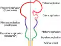
Diagram depicting the main subdivisions of the embryonic vertebrate brain. These regions will later differentiate into forebrain, midbrain and hindbrain structures.
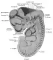
Reconstruction of peripheral nerves of a human embryo of 10.2 mm. (Label for Diencephalon is at left.)
See also
- Diencephalic syndrome
- List of regions in the human brain
References
This article incorporates text in the public domain from page 807 of the 20th edition of Gray's Anatomy (1918)
External links
- Stained brain slice images which include the "diencephalon" at the BrainMaps project
- NIF Search – Diencephalon via the Neuroscience Information Framework
This article is copied from an article on Wikipedia® - the free encyclopedia created and edited by its online user community. The text was not checked or edited by anyone on our staff. Although the vast majority of Wikipedia® encyclopedia articles provide accurate and timely information, please do not assume the accuracy of any particular article. This article is distributed under the terms of GNU Free Documentation License.